


 1 comment
Researchers at Caltech have developed a new, non-invasive technique to map brain activity using functional ultrasound technology, representing potential breakthroughs for both the study of brain function and the treatment of patients suffering from conditions such as paralysis, according to a study published Monday.
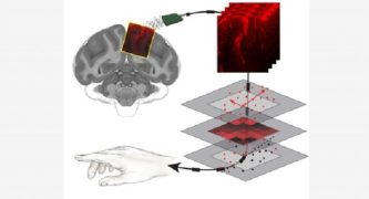
The research outlines a new method developed by scientists to use ultrasound waves, similar to those used to view fetuses, to precisely and accurately map brain activity related to movement in non-human primates, Caltech said in a written statement.
Mapping neural activity to corresponding behaviors is a major goal for neuroscientists developing brain–machine interfaces (BMIs), able to detect and interpret signals sent by the brain through brain-machine interface devices, or “devices that read and interpret brain activity and transmit instructions to a computer or machine,” according to the statement.
Such knowledge enables not only a better understanding of the brain but allows for developments such as prosthetic limbs capable of responding to the wearer’s thoughts.
“Though this may seem like science fiction, existing BMIs can, for example, connect a paralyzed person with a robotic arm; the device interprets the person’s neural activity and intentions and moves the robotic arm correspondingly,” the statement said.
But until now, there’s always been a major drawback to such technology.
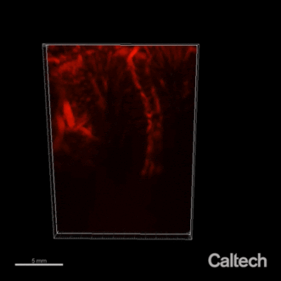
“A major limitation for the development of BMIs is that the devices require invasive brain surgery to read out neural activity,” the statement explained. “But now, a collaboration at Caltech has developed a new type of minimally invasive BMI to read out brain activity corresponding to the planning of movement. Using functional ultrasound (fUS) technology, it can accurately map brain activity from precise regions deep within the brain at a resolution of 100 micrometers. The size of a single neuron is approximately 10 micrometers.”
Existing non-invasive techniques to scan brain activity, such as functional magnetic resonance imaging, or fMRI, “can image the entire brain but require bulky and expensive machinery,” according to the Caltech statement. “Electroencephalography (EEGs) does not require surgery but can only measure activity at low spatial resolution.”
The research paper, which was published Monday in the journal Neuron, resulted from a collaboration between the labs of Caltech neuroscience professor Richard Andersen of the Tianqiao and Chrissy Chen Institute for Neuroscience and chemical engineering professor Mikhail Shapiro and affiliated faculty members in the Chen Institute, according to Caltech.
Co-first authors on the paper are Andersen lab postdoctoral fellow Sumner Norman, former Anderson lab postdoctoral scholar Vasileios Christopoulos and former Shapiro lab postdoctoral fellow David Maresca.
“When a part of the brain becomes more active, there’s an increase in blood flow to the area,” Shapiro said. “ A key question in this work was: If we have a technique like functional ultrasound that gives us high-resolution images of the brain’s blood flow dynamics in space and over time, is there enough information from that imaging to decode something useful about behavior?
“The answer is yes. This technique produced detailed images of the dynamics of neural signals in our target region that could not be seen with other non-invasive techniques like fMRI,” he said. “We produced a level of detail approaching electrophysiology, but with a far less invasive procedure.”
Among other applications, the discovery has major potential to help treat patients who have lost movement due to neurological injury or disease, Norman said.
“Invasive forms of brain–machine interfaces can already give movement back to those who have lost it due to neurological injury or disease,” she said. “Unfortunately, only a select few with the most severe paralysis are eligible and willing to have electrodes implanted into their brain. Functional ultrasound is an incredibly exciting new method to record detailed brain activity without damaging brain tissue.”
“We pushed the limits of ultrasound neuroimaging and were thrilled that it could predict movement,” Norman added. “What’s most exciting is that fUS is a young technique with huge potential — this is just our first step in bringing high performance, less invasive BMI to more people.”
The new technique is capable of delivering results 10 times more sensitive than an MRI, Maresca said.
While the recent research involved non-human primates, a collaboration is in the works with USC to study the new technology with human volunteers who have had a piece of skull removed due to traumatic brain injuries, according to Caltech,
“Because ultrasound waves can pass unaffected through these ‘acoustic windows,’ it will be possible to study how well functional ultrasound can measure and decode brain activity in these individuals,” the statement said.
The research paper can be found online at https://www.cell.com/neuron/fulltext/S0896-6273(21)00151-3#secsectitle0015.
 Get our daily Pasadena newspaper in your email box. Free.
Get all the latest Pasadena news, more than 10 fresh stories daily, 7 days a week at 7 a.m.